|
|
1.IntroductionIn the last few years, many studies have been done on the electromagnetic properties of nanostructures, particularly on the interesting applications available from their use. The extraordinary optical properties of such structures derive from the strong local electromagnetic field enhancement: the so-called localized surface plasmon resonance (LSPR). Typically, the LSPR phenomenon can be described as the resonant collective oscillation of electrons, stimulated by an incident electromagnetic wave. The resonance condition is set up when the electromagnetic wave frequency matches the natural frequency of the oscillating electrons. In addition to the LSPR, a high localization of the electromagnetic field in the neighborhood of the nanostructure is obtained. As a result, the interaction between the electromagnetic field and the surrounding dielectric environment is restricted to a small area.1 For this reason, these structures are very sensitive to any change in the local index of refraction, which causes the resonance conditions of the surface plasmon waves to modify. Such characteristics allow these structures to be suitable for several application fields, such as optical2 and photonic ones,3 biochemical sensing and detection,4,5 protein analysis,6,7 cell membrane function,8 biomedical applications,9,10 and food quality analysis.11 They can also be used as contrast agents in cellular and biological imaging.12,13 The nanoparticle effectiveness as a sensing or therapeutic tool depends on its optical properties: for instance, a high scattering cross-section is crucial for light-scattering microscopy-based imaging or sensing applications.14 On the other hand, photothermal therapy requires a high nanoparticle absorption cross-section with low scattering losses for superficial and deep tumor treatment.15 The existing literature has mainly focused on the experimental evaluation of the nanoparticle electromagnetic characteristics, but it is almost silent on the development of proper analytical models and design methods describing their resonant behavior. It is known that the analytical closed-form electromagnetic solution exists only for a restricted number of particle geometries, such as sphere and cylinder,16 cube,17 disc and needle.18 For any other arbitrary shapes, the electromagnetic solution is given by numerical approaches.19 To describe the electromagnetic behavior of the nanoparticles, it is useful to study the electromagnetic extinction cross-section properties in terms of scattering and absorption. Separate evaluation of the absorption and scattering phenomenon is crucial to understand why certain structures are preferred to others for specific applications. In order to describe the nanoparticle electromagnetic behavior using closed-form formulas, it is fundamental to correlate the electromagnetic properties, in terms of extinction cross-section, with the structural and geometrical parameters. The aim of this article is to find out the analytical relation between the nanoparticle electromagnetic properties and its geometrical parameters. In particular, this research attempts to present a new design method for ellipsoidal nanoparticles with the desired electromagnetic properties, in order to satisfy specific requirements. In the fabrication of nanorod particles, we have chosen the ellipsoidal geometry, as it represents a good approximation for the realistically obtained shapes. This article is structured as follows:
2.Analytical Models and Electromagnetic Properties of Ellipsoidal NanoparticlesThe structure under study consists of a metallic resonating inclusion embedded in a dielectric environment. Such a nanoparticle has a specific resonant wavelength in terms of magnitude, amplitude width, and position, depending on its geometrical characteristics (such as shape and dimensions) and on the metal optical properties. An electromagnetic source excites the nanostructure, and its electromagnetic properties are revealed in terms of absorption and scattering cross-section. To be more precise, when an impinging electromagnetic wave with an appropriate incident wavelength illuminates a metallic nanoparticle, free electrons of metal structure start oscillating collectively. Such oscillations lead to the propagation of strong surface waves.20,21 The resonant optical properties of nanoparticles can be studied, starting with their polarizability expressions. It is well known22 that the polarizability of an arbitrary shaped particle can be expressed as where is the particle volume, is the surrounding dielectric environment permittivity, is the inclusion dielectric permittivity, and is the depolarization factor. The polarizability value strongly depends on the nanoparticle geometry, particularly on its size, shape, inclusion composition, and the surrounding dielectric environment refractive index. Assuming that the inclusion permittivity can be written as , the dipolar polarizability is maximized (in other words, the nanoparticle is at its resonance condition) when Therefore, the factor of a nanoparticle plays a crucial role in the polarizability’s resonant behavior for the enhancement of LSPR strength. In order to study the nanoparticle electromagnetic properties in terms of scattering and absorption cross-section, the following assumptions must be made:
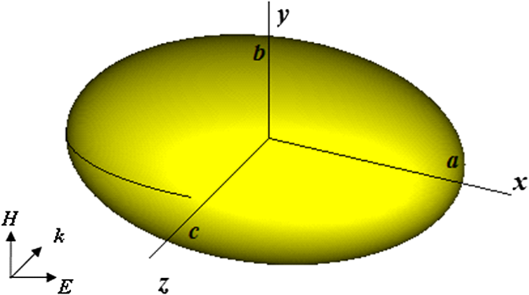
Under such conditions, we can relate the nanoparticle macroscopic electromagnetic behavior to its polarizability. In this study, the structure is excited by an electromagnetic plane wave with the electric field parallel (and the propagation vector perpendicular) to the nanoparticle principal axis, as depicted in Fig. 1, where the single particle is shown. In particular, in this study, the nanoparticles are triaxial ellipsoids with , where , , and are the semi-major axes aligned along the coordinate axes (, , ) (Fig. 1). In this paper, the following is true:
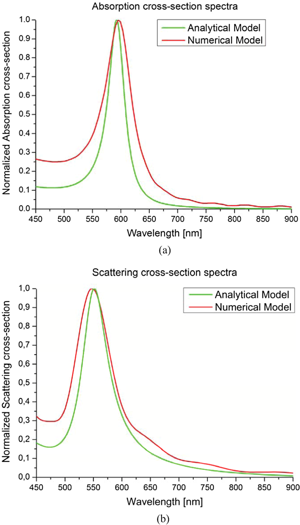
2.1.Analytical Models of Ellipsoidal NanoparticlesIn this section, the analytical models of ellipsoidal nanoparticles that link the electromagnetic nanoparticle properties to their geometrical and structural parameters are presented in order to describe their resonant behavior. Starting from the findings of Ref. 24, it is possible to develop new analytical closed-form formulas for the scattering () and absorption () cross-section of the aforementioned particles. The general corresponding expressions read, respectively, where is the wavenumber, is the wavelength, and is the refractive index of the surrounding dielectric environment. For oblate and prolate ellipsoids, several closed-form formulas for the depolarization factor can be found, while for triaxial ellipsoids, a closed formula for the depolarization factor does not exist. Typically, such factors can be written in terms of tabulated functions.25 In this study, a new closed-form analytical formula for the triaxial case is obtained. Considering the electric field polarization of the impinging plane wave and the particle geometry, the absorption and scattering cross-section formulas follow: where for triaxial ellipsoids () readswhere is the complete elliptic integral of the second kind. It is worth noting that if the triaxial ellipsoid degenerates into a prolate/oblate ellipsoid or sphere, the depolarization factor coincides with the classical formulas existing in the literature (e.g., Ref. 25). In Fig. 2, a comparison among analytical and numerical models for absorption and scattering cross-section spectra of gold triaxial ellipsoids is presented.2.2.Comparison Among Analytical, Numerical, and Experimental ResultsUsing the proposed analytical model, it is possible to describe the nanoparticle resonant behavior in order to predict its electromagnetic properties. In this subsection, the analytical results are compared to the numerical ones, performed by the finite-integration commercial code CST Studio Suite (CST, Framingham, MA), and to the experimental values of silver ellipsoids26 and gold nanorods,27 for several geometrical parameter values, as shown in Table 1. The surrounding dielectric medium is considered to be water with a complex refractive index at all wavelengths. In order to verify the quality of the proposed models, the relative errors were calculated, and they are listed in Table 1, where it is clear that a good agreement among all three results was achieved. Table 1Comparison of resonant wavelengths for triaxial ellipsoid: analytical, numerical, and experimental values (silver ellipsoids:26 b=55 nm, c=50 nm; gold nanorods:27 w is the nanorod thickness and l is the nanorod length).
2.3.Electromagnetic Properties Analysis and Sensitivity PropertiesStarting from the proposed polarizability analytical model, it is possible to predict how the geometrical parameters, the metal electromagnetic properties, and the surrounding dielectric environment permittivity affect the nanoparticle resonant behavior in terms of position, magnitude, and bandwidth. The inclusion permittivity can be expressed as . As the resonant behavior of the nanoparticle is reached when the denominator goes to zero in both its real and imaginary part [see Eq. (2)] and starting from the general polarizability expression,28 the resonant condition reads: By solving Eq. (8) with respect to and by inserting the proposed depolarization factor [Eq. (7)], we can find the corresponding solution for the inclusion permittivity for which the polarizability of the overall structure rises to infinity (which is related to the nanoparticle resonant behavior): Exploiting Eq. (9), it is possible to tune the nanostructure resonance by changing its geometrical parameters in order to coincide with the spectral absorption characteristics of the sample under study. Such a condition is necessary for specific absorption measurements. In addition, in this section, we analyze the designed nanoparticle sensitivity properties to optimize the single inclusion for sensing applications. The sensitivity is commonly defined as expressed in nm/RIU (), where is the wavelength shift and is the refractive index variation. In Table 2, we report the sensitivity values for the inclusions considered in this paper. In particular, analytical, numerical, and experimental values29 of sensitivity are compared.Table 2Comparison of the sensitivity values for the inclusions considered.
A test material surrounding the nanoparticle with a varying refractive index in the range of 1 to 3 was used. As shown in Fig. 3, the sensitivity is sufficiently constant over the range considered, as confirmed by analytical results. A good agreement among analytical, numerical, and experimental sensitivity values was obtained. Fig. 3Variation of the resonant wavelength as a function of the refractive index of the surrounding medium for the nanoparticle dimensions reported in Table 2. 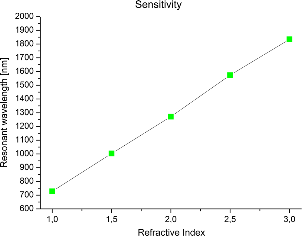 3.ConclusionsIn this paper, a study on metallic nanoparticles electromagnetic modeling in the infrared and optical frequency regime was presented. The considered structures were triaxial ellipsoidal nanoparticles, embedded in a surrounding dielectric environment. In this regard, the electromagnetic properties of such structures, in terms of extinction cross-section (absorption and scattering), were evaluated by the use of a new analytical model. Then the proposed model was compared to the results obtained by full-wave simulations and to the experimental values. A good agreement among analytical, numerical, and experimental results was achieved. The ability to manipulate and control the electromagnetic phenomenon by exploiting the proposed analytical model on the nanometer scale opens up the possibility of using such structures for sensing applications. For this reason, the nanoparticle sensitivity was studied. Sensitivity values obtained from the proposed analytical model were verified by full-wave simulations and compared to the experimental results, showing good agreement. ReferencesY.-F. Chauet al.,
“Localized resonance of composite core-shell nanospheres, nanobars, and nanospherical chains,”
Prog. Electromag. Res., 28 183
–199
(2011). http://dx.doi.org/10.2528/PIERB10102705 PELREX 1043-626X Google Scholar
J. B. Pendry,
“Playing tricks with light,”
Science, 285
(5434), 1687
–1688
(1999). http://dx.doi.org/10.1126/science.285.5434.1687 SCIEAS 0036-8075 Google Scholar
S. A. Maieret al.,
“Local detection of electromagnetic energy transport below the diffraction limit in metal nanoparticle plasmon waveguides,”
Nat. Mater., 2
(4), 229
–232
(2003). http://dx.doi.org/10.1038/nmat852 NMAACR 1476-1122 Google Scholar
J. C. Ribohet al.,
“A nanoscale optical biosensor: real-time immunoassay and nanoparticle adhesion,”
J. Phys. Chem. B, 107
(8), 1772
–1780
(2003). http://dx.doi.org/10.1021/jp022130v JPCBFK 1520-6106 Google Scholar
J. J. Storhoffet al.,
“DNA-directed synthesis of binary nanoparticle network materials,”
J. Am. Chem. Soc., 120
(48), 12674
–12675
(1998). http://dx.doi.org/10.1021/ja982721s JACSAT 0002-7863 Google Scholar
E. M. Larssonet al.,
“Sensing characteristics of NIR-localized surface plasmon resonances in gold nanoring for application as ultrasensitive biosensors,”
Nano. Lett., 7
(5), 1256
–1263
(2007). http://dx.doi.org/10.1021/nl0701612 NALEFD 1530-6984 Google Scholar
R. Bukasovet al.,
“Probing the plasmonic near-field of gold nanocrescent antennas,”
ACS Nano, 4
(11), 6639
–6650
(2010). http://dx.doi.org/10.1021/nn101994t 1936-0851 Google Scholar
W. J. Galushet al.,
“A nanocube plasmonic sensor for molecular binding on membrane surfaces,”
Nano Lett., 9
(5), 2077
–2082
(2009). http://dx.doi.org/10.1021/nl900513k NALEFD 1530-6984 Google Scholar
N. L. RosiC. A. Mirkin,
“Nanostructures in biodiagnostics,”
Chem. Rev., 105
(4), 1547
–1562
(2005). http://dx.doi.org/10.1021/cr030067f CHREAY 0009-2665 Google Scholar
J. Chenet al.,
“Gold nanocages: engineering the structures for biomedical applications,”
Adv. Mater., 17
(18), 2255
–2261
(2005). http://dx.doi.org/10.1002/(ISSN)1521-4095 ADVMEW 0935-9648 Google Scholar
H. M. Hiepet al.,
“A localized surface plasmon resonance-based immunosensor for the detection of casein in milk,”
Sci. Tech. Adv. Mat., 8
(4), 331
–338
(2007). http://dx.doi.org/10.1016/j.stam.2006.12.010 STAMCV 1468-6996 Google Scholar
D. Yelinet al.,
“Multiphoton plasmon-resonance microscopy,”
Opt. Exp., 11
(12), 1385
–1391
(2003). http://dx.doi.org/10.1364/OE.11.001385 OPEXFF 1094-4087 Google Scholar
K. Sokolovet al.,
“Real-time vital optical imaging of precancer using anti-epidermal growth factor receptor antibodies conjugated to gold nanoparticles,”
Cancer Res., 63
(9), 1999
–2004
(2003). CNREA8 0008-5472 Google Scholar
W. Caiet al.,
“Application of gold nanoparticles in cancer nanotechnology,”
Nano. Sci. App., 1
(1), 17
–32
(2008). http://dx.doi.org/10.2147/NSA.S3788 NSAAAX 1177-8903 Google Scholar
I. H. El-SayedX. HuangM. A. El-Sayed,
“surface plasmon resonance scattering and absorption of anti-EGFR antibody conjugated gold nanoparticles in cancer diagnostics: applications in oral cancer,”
Nano. Lett., 5
(5), 829
–834
(2005). http://dx.doi.org/10.1021/nl050074e NALEFD 1530-6984 Google Scholar
A. D. Yaghjian,
“Electric dyadic Green’s functions in the source region,”
Proc. IEEE, 68
(2), 248
–263
(1980). http://dx.doi.org/10.1109/PROC.1980.11620 IEEPAD 0018-9219 Google Scholar
J. Avelinet al.,
“Electric fields in the source region: the depolarization dyadic for a cubic cavity,”
Elect. Eng., 81
(4), 199
–202
(1998). http://dx.doi.org/10.1007/BF01233270 1472-1287 Google Scholar
L. D. LandauE. M. Lifshitz, Electrodynamics of Continuous Media, 34
–85 Pergamon Press, Oxford
(1984). Google Scholar
A. Sihvola,
“Dielectric polarization and particle shape effects,”
J. Nano., 2007
(1), 1
–9
(2007). http://dx.doi.org/10.1155/2007/45090 JNOABP 1687-4129 Google Scholar
S. Kessentiniet al.,
“Selective and collaborative optimization methods for plasmonic: a comparison,”
PIERS Online, 7
(3), 291
–295
(2011). http://dx.doi.org/10.2529/PIERS100902052449 Google Scholar
P. K. JainM. A. El-Sayed,
“Plasmonic coupling in noble metal nanostructures,”
Chem. Phys. Lett., 487
(4), 153
–164
(2010). http://dx.doi.org/10.1016/j.cplett.2010.01.062 CHPLBC 0009-2614 Google Scholar
C. BohrenD. Huffmann, Absorption and Scattering of Light by Small Particles, 57
–81 John Wiley, New York
(1983). Google Scholar
P. B. JohnsonR. W. Christy,
“Optical constants of the noble metals,”
Phys. Rev. B, 6
(12), 4370
–4379
(1972). http://dx.doi.org/10.1103/PhysRevB.6.4370 PRBMDO 1098-0121 Google Scholar
J. G. Van Bladel, Electromagnetic Fields, 77
–124 John Wiley & Sons, Hoboken
(2007). Google Scholar
A. Sihvola, Electromagnetic Mixing Formulas and Applications, The Institution of Engineering and Technology, London
(2008). Google Scholar
B. J. Wileyet al.,
“Synthesis and optical properties of silver nanobars and nanorice,”
Nano Lett., 7
(4), 1032
–1036
(2007). http://dx.doi.org/10.1021/nl070214f NALEFD 1530-6984 Google Scholar
K. Uenoet al.,
“Spectral sensitivity of uniform arrays of gold nanorods to dielectric environment,”
J. Phys. Chem. C, 111
(11), 4180
–4184
(2007). http://dx.doi.org/10.1021/jp068243m 1932-7447 Google Scholar
L. La SpadaR. IovineL. Vegni,
“Nanoparticle electromagnetic properties for sensing applications,”
Adv. Nano., 1
(2), 9
–14
(2012). http://dx.doi.org/10.4236/anp.2012.12002 69MTLT Google Scholar
H. Lianget al.,
“Silver nanorice structures: Oriented attachment-dominated growth, high environmental sensitivity, and real-space visualization of multipolar resonances,”
Chem. Mater., 24
(12), 2339
–2346
(2012). http://dx.doi.org/10.1021/cm3006875 CMATEX 0897-4756 Google Scholar
Biography Luigi La Spada received a bachelor’s degree, summa cum laude, in electronics engineering from the University of Roma Tre, Rome, Italy, in 2008. In 2010, he received his master’s degree, summa cum laude, in information and communication technology from the University of Roma Tre. Since 2011, he has been with the Department of Applied Electronics, University of Roma Tre, where he is working as a PhD student (scholarship winner) at the Doctoral School in Engineering in the biomedical electronics, electromagnetics, and telecommunications section. His main research interests are microwave, THz, and optical applications of complex media and metamaterials; design of miniaturized microwave, THz, and infrared sensors based on metamaterial technology; optical properties, biological and biomedical applications of plasmonic nanoparticles; and analysis and synthesis of planar metamaterials and radiating elements for sensing applications.  Renato Iovine received the first-level laurea degree summa cum laude in electronics engineering at University of Roma Tre, in December 2009. In October 2011, he received the second-level laurea degree (Laurea Magistralis) summa cum laude in bioengineering at University of Roma Tre. Now he is a PhD student (scholarship winner) in biomedical electronics, electromagnetics, and telecomunications. His main research interests are optical properties of plasmonic nanoparticles, design of nanoantennas for sensing applications, design of biomedical sensor based on miniaturized metamaterials, and biological integrated planar and conformal sensors.  Lucio Vegni received the degree in electronics engineering from the University of Rome, Rome Italy. After working at Standard Elektrik Lorenz in Stuttgart (West Germany) as an antenna designer, he joined the Istituto di Elettronica of the University of Rome, Rome, Italy, where he was involved in research work in the areas of antenna and scattering problems from 1970 to 1992. Since 1992, he has been an associate professor of electromagnetic compatibility at the University of Rome “La Sapienza,” and then he was a full professor at the Third University of Rome, Rome, Italy, where he founded the Research Laboratory in Applied Electromagnetics. His research interests are in the areas of microwave components and antennas, with particular emphasis on biological integrated planar and conformal sensors and antennas loaded with complex materials, such as chiral media and metamaterials. He is the author of more than 700 papers on complex media and metamaterials published on international journals and conference proceedings. He is a life member of IEEE. |
|||||||||||||||||||||||||||||||||||||||||||||||||||||||||||||||||||||||