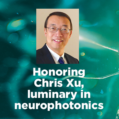
Chris Xu
Chris Xu is known for his contribution to the development and invention of three-photon microscopy, notably in a 2013 Nature Photonics paper. As a graduate student, Xu, working with Cornell's Watt Webb in Webb’s lab, originally demonstrated three-photon fluorescence imaging in 1996. Today, Xu is the IBM Professor of Engineering and director of the School of Applied and Engineering Physics at Cornell University. Read more about Chris Xu’s contributions to the field of neurophotonics on SPIE News, or read about other SPIE Luminaries.

Imaging deeper, wider, and faster (Conference Presentation)
(Proceedings of SPIE, 2020)
High-speed, large field-of-view and deep imaging with an adaptive excitation source
(Proceedings of SPIE, 2020)
Time-lens based multi-color background-free coherent anti-Stokes Raman scattering microscopy
(Proceedings of SPIE, 2019)
In vivo label-free confocal imaging of adult mouse brain up to 1.3-mm depth with NIR-II illumination
(Proceedings of SPIE, 2019)
Comparison of emission wavelengths for in vivo deep imaging of mouse brain
(Proceedings of SPIE, 2019)
Whole-brain observation in a living Drosophila brain by three-photon excitation at 1300-nm (Conference Presentation)
(Proceedings of SPIE, 2018)
In vivo three-photon imaging of deep cerebellum
(Proceedings of SPIE, 2018)
Comparison of excitation wavelengths for in vivo deep imaging of mouse brain
(Proceedings of SPIE, 2018)
Third harmonic generation imaging of intact human cerebral organoids to assess key components of early neurogenesis in Rett Syndrome (Conference Presentation)
(Proceedings of SPIE, 2017)
In vivo three-photon activity imaging of GCaMP6-labeled neurons in deep cortex and the hippocampus of the mouse brain
(Proceedings of SPIE, 2017)
Multi-wavelength time-lens source and its application to nonresonant background suppression for coherent anti-Stokes Raman scattering microscopy
(Proceedings of SPIE, 2017)
Nonlinear adaptive optics: aberration correction in three photon fluorescence microscopy for mouse brain imaging
(Proceedings of SPIE, 2017)
Direct visualization of functional heterogeneity in hepatobiliary metabolism
(Proceedings of SPIE, 2017)
Multiphoton gradient index endoscopy for evaluation of diseased human prostatic tissue ex vivo
(J. of Biomedical Optics, 2014)
Two-photon fluorescence imaging of intracellular hydrogen peroxide with chemoselective fluorescent probes
(J. of Biomedical Optics, 2013)
Special Section Guest Editorial: Multiphoton Microscopy: Technical Innovations, Biological Applications, and Clinical Diagnostics
(J. of Biomedical Optics, 2013)
High resolution, large field-of-view endomicroscope with optical zoom capability
(Proceedings of SPIE, 2013)
Multi-color femtosecond source for simultaneous excitation of multiple fluorescent proteins in two-photon fluorescence microscopy
(Proceedings of SPIE, 2013)
Spectroscopic SRS imaging with a time-lens source synchronized to a femtosecond pulse shaper
(Proceedings of SPIE, 2013)
In vivo imaging of unstained tissues using a compact and flexible multiphoton microendoscope
(J. of Biomedical Optics, 2012)
Fiber delivered two-color picosecond source for coherent Raman scattering imaging
(Proceedings of SPIE, 2012)
In vivo two-photon microscopy to 1.6-mm depth in mouse cortex
(J. of Biomedical Optics, 2011)
Epifluorescence light collection for multiphoton microscopic endoscopy
(Proceedings of SPIE, 2011)
Multiphoton imaging for deep tissue penetration and clinical endoscopy
(Proceedings of SPIE, 2011)
Miniaturized fiber raster scanner for endoscopy
(Proceedings of SPIE, 2011)


